Farming
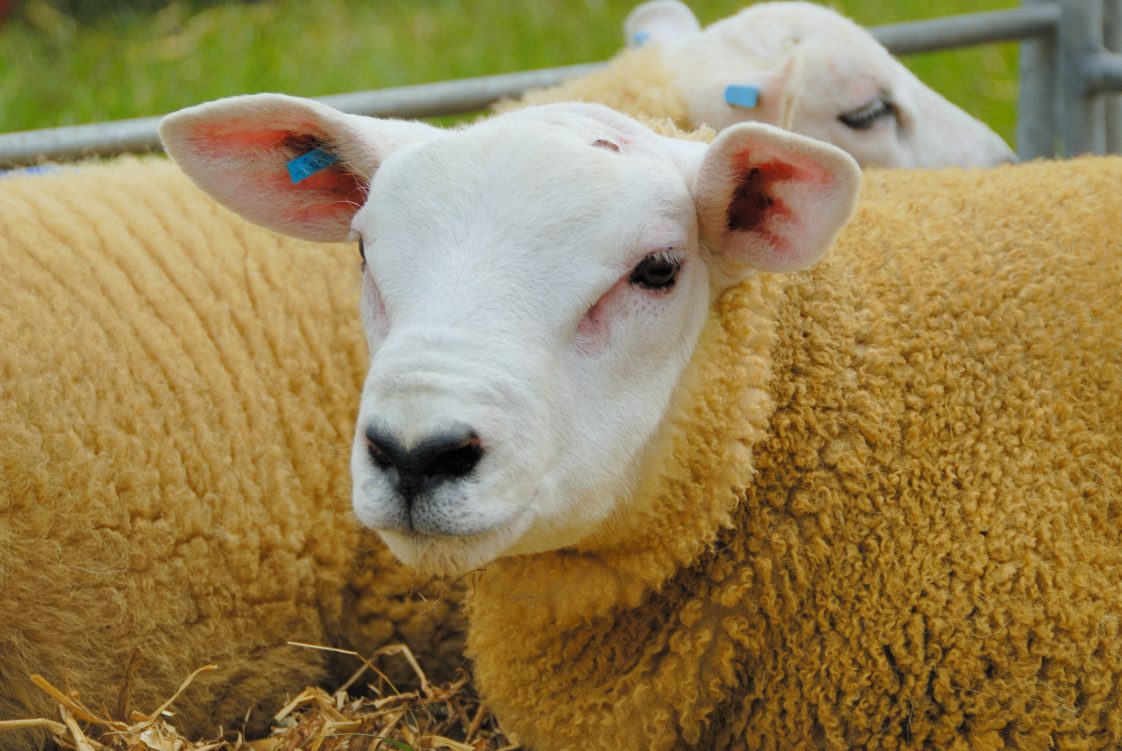
Neonatal lambs and kids are susceptible to a range of eye conditions, from common treatable issues to rare congenital defects. Among the most frequent problems is entropion, where the eyelid rolls inward and irritates the cornea, the outer layer of the eye. Another common issue is infectious conjunctivitis (pinkeye), especially in young animals kept in crowded or dusty environments.
Additionally, certain breeds are born with congenital eye anomalies, such as microphthalmia (small eyes) or anophthalmia (absence of one or both eyes). Other congenital defects include iris or retinal colobomas (missing eye tissue), aphakia (absence of the lens), and dermoids (skin-like growths on the cornea). These anomalies often occur together, such as microphthalmia with colobomas or anophthalmia with dermoids (non-cancerous cysts).
Causes and Risk Factors
Several factors contribute to the development of eye conditions in neonatal small ruminants, including genetic predisposition, infectious agents, nutritional deficiencies, and environmental influences.
- Genetics. Genetic predisposition and breeding are significant risk factors for this condition. Entropion is inherited in many flocks, and certain sheep breeds, such as Texel, Ile de France, or Charollais, exhibit higher incidences. A specific mutation in the PITX3 gene is linked to microphthalmia in Texel sheep. Breed and family history should be taken into consideration. Rams that sire lambs with entropion or congenital eye defects should be removed from the flock.
- Infectious agents. Infectious agents can lead to neonatal ocular disease. Contagious pinkeye pathogens (Chlamydophila pecorum, Mycoplasma conjunctivae, Moraxella ovis) are common in sheep and goats. Young stock in summer, dusty or poor hygiene conditions are at higher risk since dust, flies, and overcrowding help spread pathogens. Additionally, systemic infections such as Listeriosis in pregnant dams, often caused by bacterial contamination from spoiled feed, can lead to fetal eye conditions such as uveitis or optic nerve damage in neonates.
- Nutritional deficiencies. Insufficient vitamin A in the dam’s diet can result in fetal ocular malformations, including microphthalmia and colobomas.
- Environment and management. Environmental and management factors also contribute to risk. Birth trauma or injury during penning, such as horn thrusts or abrasive bedding, can damage a neonate’s eye. Sunlight and forage contaminants, including plant toxins, can irritate or harm the cornea. Poor sanitation may also introduce chemical irritants. Overall, risk factors include genetics (heredity), maternal health or nutrition, infectious exposure, and the perinatal environment.
Clinical Signs and Symptoms
The clinical presentation varies depending on the condition.
- Entropion. An inward-rolled eyelid (usually the lower eyelid) is common and often hereditary. Affected lambs or kids exhibit epiphora (overflow tearing), blepharospasm (squinting), and persistent ocular discharge. The eyelid rim is rolled inward, causing eyelashes and hair to rub the cornea. In advanced cases, keratitis or corneal ulcers may develop, which can lead to corneal opacity and potential blindness. Typically, one or both eyes are involved from birth or within the first week.
- Infectious keratoconjunctivitis (pinkeye). Bacterial keratoconjunctivitis is caused by Chlamydia or Mycoplasma (seasonal, contagious). Signs include blepharospasm, photophobia, and purulent/mucopurulent ocular discharge. The conjunctiva becomes red and swollen, and epiphora and eyelid crusting are common. Corneal lesions, such as ulcers or white opacities, may appear. Affected neonates may also exhibit signs of discomfort, such as depression, although systemic signs are uncommon in simple eye infections.
- Neonatal conjunctivitis (non-specific). Symptoms may include tearing and mild discharge, with the condition resembling pinkeye but possibly less severe or chemical in nature (e.g., from high ammonia levels). If infectious, the pathology overlaps with infectious keratoconjunctivitis.
- Congenital anomalies. Microphthalmia, anophthalmia, colobomas, cataracts, aphakia, and dermoids (rare and often genetic or teratogenic) are common congenital anomalies. These conditions are visible at birth or shortly after. Microphthalmic animals have abnormally small eyes, while anophthalmic animals have no eyes at all. The corneas may appear opaque or malformed, and the lenses may be absent (aphakia) or cataractous. Animals with severe congenital defects are blind in the affected eyes. Dermoids appear as fleshy, hair-covered plaques on the cornea or conjunctiva, while colobomas result in keyhole-shaped pupils or iris defects. These anomalies do not cause pain but lead to vision loss and may result in growth retardation, as affected animals may fail to thrive.
Diagnostic Approaches
Diagnosing neonatal eye conditions begins with a thorough clinical examination. Fluorescein dye tests are useful in detecting corneal ulcers, while careful observation of the eyelids helps identify entropion.
For conjunctivitis, samples taken from the conjunctiva, such as swabs or scrapings, can be tested for infectious agents using microscopy or molecular methods. In some cases, tear flow tests or intraocular pressure measurements may be conducted if other conditions, such as glaucoma, are suspected.
Imaging techniques like ocular ultrasonography help assess internal eye structures when visual examination is not possible due to opacity or inflammation. For congenital defects, genetic testing can confirm inherited eye conditions, while post-mortem histological analysis may be required to diagnose rare or complex developmental abnormalities.
Treatment Options
- Entropion: Prompt correction is crucial to prevent corneal ulceration. For mild cases, simply everting the lid and applying lubricant or antibiotic ointment can suffice. A common field therapy is to inject a mild irritant (e.g., long-acting penicillin or hypertonic saline) into the eyelid to create a scar that rolls the lid outward. In more severe or persistent cases, surgical intervention is needed: eyelid tacking (using sutures, staples, or clips) or a wedge resection (eyelid excision) permanently corrects the eyelid position. After any procedure, topical antibiotics and anti-inflammatory drops are used to prevent infection and pain. High success rates have been reported using irritant injection methods.
- Infectious conjunctivitis (pinkeye): Antimicrobial therapy is the mainstay. Topical tetracycline ointment applied daily can control many cases. Systemic antibiotics are used in outbreaks or severe cases. Although no drugs are specifically labeled for sheep and goat keratoconjunctivitis, extra-label use of oxytetracycline injections or macrolides, such as tulathromycin, is common. Infected animals should be isolated and have any soiling material cleaned. Eye patches or protective hoods can also be used to shield ulcers from light and flies. For deep or non-healing corneal ulcers, a third-eyelid flap or conjunctival graft may be performed by a specialist to promote healing. Topical atropine can relieve ciliary spasm (pain) in severe cases, and nonsteroidal anti-inflammatory drugs (NSAIDs) provide systemic pain relief.
- Congenital defects: Unfortunately, there is no curative treatment for the most severe congenital anomalies. Bilateral anophthalmia or profound microphthalmia usually warrants humane euthanasia soon after birth. Unilateral defects may allow one functional eye, so the animal can be managed if it can feed and thrive. Some anterior segment anomalies (small cataracts, incomplete colobomas) can be monitored; surgical lens removal or corneal grafting is rarely performed in farm animals. For inherited conditions, the main treatment is breeding management, which selects against carriers by using genetic tests.
Prevention and Management
Preventive strategies for neonatal eye conditions focus on effective management practices and genetic selection.
- Genetic selection. Regularly inspect newborns to detect conditions like entropion early, allowing for timely intervention. Animals with hereditary eye defects should be excluded from breeding to reduce the prevalence of these conditions in future generations.
- Maternal nutrition. Proper nutrition, particularly adequate vitamin intake during pregnancy, is essential in preventing fetal ocular malformations. Ensuring clean and dry birthing areas also helps minimize the risk of infection in neonates.
- Environmental management. In cases of infectious conjunctivitis, isolation of affected animals, routine cleaning of equipment, and management of insect vectors can help prevent outbreaks. Adequate space, shade, and good ventilation should be provided to reduce stress and support overall eye health.
- Vaccination. Although not commonly used for most ocular conditions, vaccination may be an option if specific pathogens are identified. Genetic testing can help identify and eliminate inherited eye diseases from breeding populations.
Prognosis
The prognosis for neonatal eye conditions depends on the type and severity of the disorder and how quickly it is diagnosed and treated.
- Entropion: With prompt treatment, the prognosis is excellent. Most affected animals recover fully, but untreated entropion can lead to permanent corneal damage and blindness. Some lambs may require multiple treatments if the condition recurs.
- Infectious conjunctivitis (pinkeye): The prognosis is generally good, with most animals recovering within a few weeks. However, recurrent or untreated infections may result in lasting eye damage, such as cataracts or corneal scarring.
- Congenital anomalies: These often carry a poor prognosis, particularly in cases of bilateral blindness or major malformations. Affected animals may fail to thrive or survive. However, animals with one affected eye or minor defects may adapt and live productive lives, depending on management practices.
Early identification and intervention greatly improve outcomes for neonatal eye conditions, making routine checks in the first days of life essential for ensuring the long-term health and productivity of affected lambs and kids.
Standard Prevention and Management
Prevention focuses on management and genetics. For entropion, routine neonatal checks should ensure eyelids are normal. Lambs and kids with entropion are often treated or culled, and rams that sire affected offspring should be removed from the breeding pool. Good hygiene, fly control, and early detection limit conjunctivitis outbreaks. Genetic counseling and screening programs help reduce the prevalence of inherited ocular defects.